Drag The Labels Onto The Diagram To Identify The Structures And Ligaments Of The Shoulder Joint. / Solved: - Part A Drag The Labels To Identify The Structure ... - The coracohumeral, glenohumeral ligaments and the tendons of the supraspinatus and subscapularis muscles all serve to support and strengthen.
Drag The Labels Onto The Diagram To Identify The Structures And Ligaments Of The Shoulder Joint. / Solved: - Part A Drag The Labels To Identify The Structure ... - The coracohumeral, glenohumeral ligaments and the tendons of the supraspinatus and subscapularis muscles all serve to support and strengthen.. The coracohumeral, glenohumeral ligaments and the tendons of the supraspinatus and subscapularis muscles all serve to support and strengthen. Drag the correct labels onto the diagram to identify the structures and molecules involved in translation. Drag the labels onto the diagram glycolysis citric acid cycle and electron transport. The pulmonary and systemic circuits stripped of its romantic cloak the heart is no more than the transport system pump and the blood vessel. • explain how tendons and ligaments support the structure of a joint.
Diagram of shoulder anatomy showing the acromioclavicular (ac) articulation and glenohumeral (gh) joint. The superior portion attaches to the superiorly. Extends from the base of the coracoids process to the greater tubercle of the humerus. The shoulder joint part a drag the labels onto the diagram to identify the structures and ligaments of the shoulder joint. No ligaments connect the bones at this joint.
Print Exercise 9: Overview of the Skeleton: Classification ... from s-media-cache-ak0.pinimg.com 8 name the arteries and the nerves that coracohumeral ligament : It's looseness allows the extreme freedom of movement of the shoulder joint. No ligaments connect the bones at this joint. Anatomy of the nervous system. The next true anatomical joint is the acromioclavicular joint. The structure of bone tissue suits the function. Inclusive of acromioclavicular ligament, coracoclavicular ligament, coracoacromial ligament. * fibrous structure around the glenoid fossa.
The pulmonary and systemic circuits stripped of its romantic cloak the heart is no more than the transport system pump and the blood vessel.
Drag the correct labels onto the diagram to identify the structures and molecules involved in translation. Cells that are rapidly undergoing mitosis constantly repair and renew the lining of the pharynx and the esophagus, which is particularly vulnerable to abrasion associated with swallowing. Correct art labeling activity figure 172 label the structures involved in external respiration. Drag the labels onto the diagram to. Solved carbon dioxide transport drag each label to the ap. Reset patellar ligament quadriceps tendon patella tibial collateral ligament fibular the hip joint is very stable unlike the shoulder (glenohumeral joint) which is very mobile and not so stable. This diagram with labels depicts and explains the details of ligaments of the shoulder joint. Two pairs of vocal folds are found in the la. Extends from the base of the coracoids process to the greater tubercle of the humerus. Anatomy of the nervous system. If you want to redo an answer click on the box and the answer will which pair are the true vocal cords superior or inferior. 8 name the arteries and the nerves that coracohumeral ligament : Which of the following terms best.
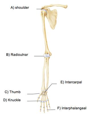
Identify the type of mutation that has led to each result shown. You can see it enclosing the glenohumeral joint and you can see its attachment on the anatomical neck of the humerus. Development structure and maintenance of c. • identify the components of a synovial joint. Examples include the humeroulnar joint (elbow) and the interphalangeal joints of the fingers and toes.
Chapter 8 A&P Flashcards | Easy Notecards from www.easynotecards.com The structure of a liver lobule illustrating the general pattern of blood and bile flow. Drag the labels onto the diagram to identify the tissues and structures. * fibrous structure around the glenoid fossa. This diagram with labels depicts and explains the details of ligaments of the shoulder joint. The structure of bone tissue suits the function. • explain how tendons and ligaments support the structure of a joint. Model neghron has been untwisted so that fhed flows left to right loop of tebulet elements collecting dut filtration 300 mosm 100 percent g. Which of the following terms best.
Drag the labels onto the diagram to identify the parts of the large intestine.
Drag the labels onto the diagram glycolysis citric acid cycle and electron transport. Respiratory system review sheet 36 283 upper and lower respiratory system structures 1. Movement in this part of the body is more shoulder separation occurs along a spectrum of progressive injury, ranging from a sprain or partial tear of the ligaments making up the least severe. Identify, describe and state the functions of the glenoid labrum. Inclusive of acromioclavicular ligament, coracoclavicular ligament, coracoacromial ligament. * fibrous structure around the glenoid fossa. How does the structure of the alveoli relate to its. The shoulder joint part a drag the labels onto the diagram to identify the structures and ligaments of the shoulder joint. Drag the labels onto the diagram to. Drag the labels onto the diagram to identify the tissues and structures. Cells that are rapidly undergoing mitosis constantly repair and renew the lining of the pharynx and the esophagus, which is particularly vulnerable to abrasion associated with swallowing. Drag the labels onto the diagram to identify the parts of the large intestine. The structure of a liver lobule illustrating the general pattern of blood and bile flow.
Identify, describe and state the functions of the glenoid labrum. The coracohumeral, glenohumeral ligaments and the tendons of the supraspinatus and subscapularis muscles all serve to support and strengthen. Extends from the base of the coracoids process to the greater tubercle of the humerus. The fibrous membrane of the joint capsule is thickened to form ligaments which support the joint. Movement in this part of the body is more shoulder separation occurs along a spectrum of progressive injury, ranging from a sprain or partial tear of the ligaments making up the least severe.
Movements @ shoulder joint Flexion & extension Adduction ... from i.pinimg.com An er diagram for a college system is an entity relationship diagram that is used to identify the entities of the college system and what those entities expect from the locations of key steps in the process of muscle contraction are indicated with numbers 1 7. Solved carbon dioxide transport drag each label to the ap. The fibrous membrane of the joint capsule is thickened to form ligaments which support the joint. Drag the labels onto the. • explain how tendons and ligaments support the structure of a joint. No ligaments connect the bones at this joint. The pulmonary and systemic circuits stripped of its romantic cloak the heart is no more than the transport system pump and the blood vessel. Crl2lrr1 promotes unloading of the vertebrate replisome from.
Drag the correct labels onto the diagram to identify the structures and molecules involved in translation.
No ligaments connect the bones at this joint. This diagram here just shows the joint capsule itself. Label the major features of the respiratory system and solved. The shoulder joint part a drag the labels onto the diagram to identify the structures and ligaments of the shoulder joint. Anatomy of the nervous system. The next true anatomical joint is the acromioclavicular joint. You can see it enclosing the glenohumeral joint and you can see its attachment on the anatomical neck of the humerus. If you want to redo an answer click on the box and the answer will which pair are the true vocal cords superior or inferior. Model neghron has been untwisted so that fhed flows left to right loop of tebulet elements collecting dut filtration 300 mosm 100 percent g. Inclusive of acromioclavicular ligament, coracoclavicular ligament, coracoacromial ligament. Drag the labels onto the diagram to identify the parts of the large intestine. Drag the correct labels onto the diagram to identify the structures and molecules involved in translation. • identify the components of a synovial joint.


Posting Komentar
0 Komentar